Eyes
The eyes and how they work
The eyes and how they work
Seeing is a complex process. The brain is responsible for vision more than hearing, tasting, touching and smelling. In this article we will explain the anatomy of the eye and how we see.
Here’s an overview of how the visual system works.
Light passes through the cornea, a dome-shaped structure. The cornea refracts light and helps the eye focus.
Light passes through the lens. Together with the cornea, the lens focuses light onto the retina at the back of the eye.
The optic nerve sends impulses to the brain, which processes the signal and creates an image.
To understand how this happens, let’s start by studying the anatomy of the eye.
Below is an interactive 3D model of the eye. To learn more about the eye, explore the model using the mouse pad or touch screen.
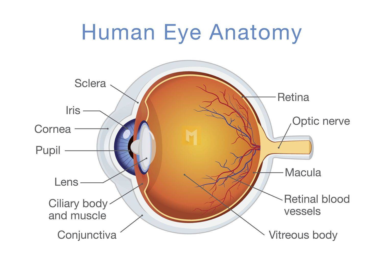
Anatomy of the eye
The side of the eye that people see is the front side. The rest are inside the eye or orbit. Muscles connected to the eyeballs allow the eyes to move in the direction of a person’s gaze.
There are three main types of eye tissue:
- A refraction grid that focuses light
- Light-sensitive fabric
- A supportive network
We’ll explore each of these types below.
Network bias
A light-focusing refraction grid is created in the light-sensitive grid, which gives a clear and sharp image. If the tissue is disordered, disorganized, or damaged, vision can become blurred.
Refractory tissue is:
University student
It is a dark spot in the center of the colored part of the eye.
When exposed to bright light, the pupil constricts to protect the sensitive retina from damage. It grows in low light. This allows the eye to perceive the most light.
Iris
This part of the eye is colored. It contains muscles that control the size of the pupil and the amount of light that reaches the retina. In this sense, it is similar to the aperture of a camera.
Lens
After passing through the pupil, the light reaches the lens. It is a transparent curved structure. The lens can change shape and help focus light on the retina. As the lens ages, it becomes harder and more flexible, making it harder to focus.
Ciliary muscle
It is a muscular ring attached to the lens. This process is called adaptation.
Cornea
The cornea is a transparent, dome-shaped layer that covers the pupil, iris, and anterior chamber of the eye. This section is the fluid-filled area between the cornea and the iris.
The cornea, like the eyelid, lid, and tear fluid, protects the eye from damage and foreign bodies such as dust. It also helps focus the eyes by letting light into the eyes.
The cornea is full of nerve endings and is very sensitive. It is the eye’s first line of defense against foreign bodies and injury. Since the cornea must be clear to refract light, there are no blood vessels inside.
Light sensitive tissue
This includes the retina and optic nerve.
The name of the retina
The retina is the inner layer of the eye. It consists of millions of reliable, light-sensitive photoreceptor cells that detect light and convert it into electrical signals. These signals are sent to the brain for processing.
The two basic photoreceptor cells are called “rods” and “cones”. When it detects light, it sends an electrical signal to the brain.
This cone is located in the center of the retina, the macula. The retina contains about 6 million cones. The fovea is a small hole in the center of the macula with a high density of cone cells and no rods.
Under normal lighting conditions, cones help people see and distinguish colors. There are different types according to color sensitivity. These are roughly as follows:
- red
- vegetable
- blue
Red and green cones are usually located in the center of the fovea and blue cones are usually outside.
Rods are usually located around the edges of the retina. Those responsible for the black and white vision are a reliable source. They can detect minimal light and see people at night.
The nerve of the eye
The optic nerve is a thick bundle of nerve fibers that carries signals from the retina to the brain. Thin retinal fibers called tube cells carry light information from the eye to the brain.
Ganglion cells protrude from the eye at a point called the optic disc. It is also called a “blind spot” because it has no rod or cone.
Different types of ganglion cells register different types of visual information. For example, some people are sensitive to contrast and movement, while others are sensitive to shape and detail. Together they transfer all the necessary information to our field of vision.
Brain
The brain integrates signals from both eyes to provide depth perception.
Signals from the eye enter the visual cortex, which processes visual information in the brain. The visual cortex integrates stimuli from both eyes into images.
The condition of the eyes
Several health conditions can affect your eyesight. These can be:
- The genetic component
- Human innate characteristics.
- age
- Other health problems
Here are some examples.
Pigmentation: Also called pigmentation, this genetic condition affects cone cells. People have trouble distinguishing certain colors.
Age-related macular degeneration: blurring of the center of the visual field. This can cause vision loss.
Amblyopia: This condition begins in childhood and is often referred to as “lazy eye.” One eye is not fully developed because the other, stronger eye is too much.
Anisocoria: This happens when the pupils are irregularly shaped. This may be harmless, but could indicate a more serious medical problem, such as a stroke.
Astigmatism: An incorrectly curved cornea or lens that prevents light from properly reaching the retina.
Cataract: Clouding of the lens. This can cause vision loss.
Chalazion: The formation of a nodule due to closing of the eyelids. It’s like a stable, but it’s not caused by an infection.
Conjunctivitis: Also called conjunctivitis, it is an infection of the conjunctiva that covers the front of the eyeball.
When to see a doctor
You should see your doctor if:
- Any sudden changes, including sudden increases in floaters
- Severe pain and redness
- Severe sensitivity to light
- Vision loss, double vision, or other vision changes
- Injuries affecting the eye or orbit.
- Green tissue is light sensitive
This includes the retina and optic nerve.
The name of La Ritain
La retin est la sofa la plus interne de l’oeil. It contains millions of light-sensitive photoreceptor cells that detect light and convert it into electrical signals. These signals are sent to the brain for treatment.
Les Deux Cellules photoreceptor primers are called “batonettes” and “cones”. Lorscur’ils detects the light, the envoy of signal electricity or servo.
The cones are located in the center of the retina, the macula. The retina contains about 6 million cones. The fovea, a small fosse in the middle of the macula, has a high density of cellular coniques and passes of baton nets.
The cones identify the people who are a voyeur in the typical conditions of eclairs and distinguish the coolers. There are different types according to color sensitivity.